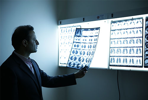
We believe an accurate and timely diagnosis of a disease is where the best course of its treatment begins. We offer an ample range of diagnostic imaging services, with our skilled staff using the latest testing equipment at out Chest Radiology Center.
Risks
The amount of radiation from a chest X-ray is lower than most people believe. We are naturally exposed to far higher levels of radiation in the environment.
Despite this, extra precautions are often taken and patients are given protective gear for surrounding organs if more than one image is required.
Do tell your doctor if you’re pregnant or might be pregnant so it is taken into account and your abdomen can be shielded for added protection.
 Results
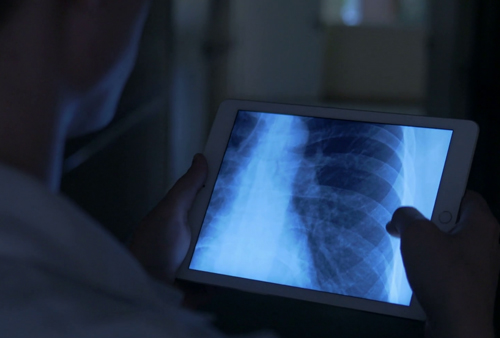
A chest X-ray generates a black-and-white image of the organs in your chest. White structures on these images are the ones which block the radiation, while the black are the ones which let the radiation pass through.
Your bones appear white because of their density. Your heart also appears as a lighter area. Lungs block very little radiation as they are filled with air sacs, so they appear as darker areas on the images.
A radiologist is a specialist in X-rays and other imaging exams, and is responsible for analyzing the images and identifying any anomalies in the chest cavity, including signs of heart problems, collection of liquid anywhere in the chest, infection, signs of growths or cancers or anything to suggest any specific other diseases.
They compile a detailed report of their findings, thus enabling the treating lung or thoracic specialist to manage the condition most effectively. The consulting doctor will discuss the results with the patient, as well as what treatments, precautions and practices are required for optimal treatment of the presenting issue.
